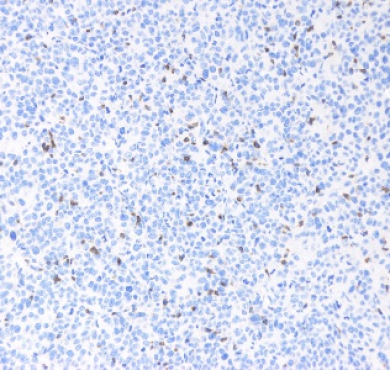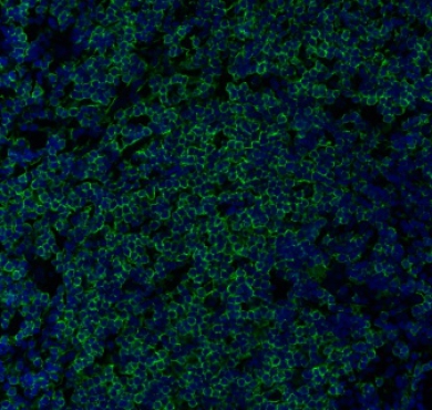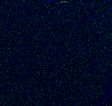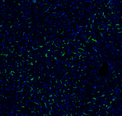
Anti-Vimentin antibody[STJ96245]
Product nameAnti-Vimentin antibodyShort DescriptionRabbit polyclonal against VimentinDescriptionRabbit polyclonal to Vimentin
Product nameAnti-Vimentin antibodyShort DescriptionRabbit polyclonal against VimentinDescriptionRabbit polyclonal to Vimentin

| Product name | Anti-Vimentin antibody |
|---|---|
| Short Description | Rabbit polyclonal against Vimentin |
| Description | Rabbit polyclonal to Vimentin. |
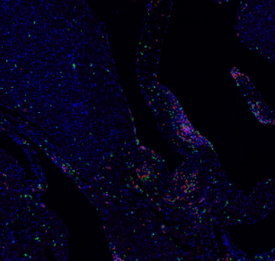
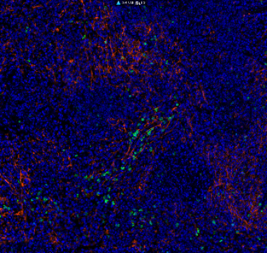
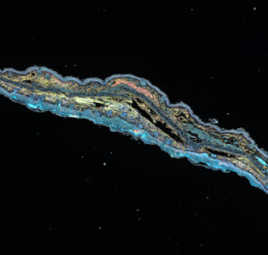
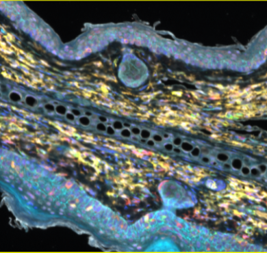
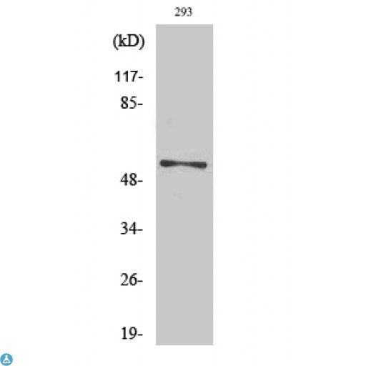
| Applications | ELISA, IF, IHC, WB |
| Dilution range | WB 1:500-1:2000 IHC 1:100-1:300 IF 1:200-1:1000 ELISA 1:10000 |
| Specificity | Vimentin Polyclonal Antibody detects endogenous levels of Vimentin protein. |
| Protein Name | Vimentin |
| Immunogen | Synthesized peptide derived from human Vimentin around the non-phosphorylation site of S83. |
| Immunogen Region | 55-105 aa |
| Storage Instruction | Store at -20°C, and avoid repeat freeze-thaw cycles. |
| Note | For Research Use Only (RUO). |
| Validated Application |
| Host | Rabbit |
|---|---|
| Clonality | Polyclonal |
| Reactivity | Human, Mouse, Rat |
| Conjugation | Unconjugated |
| Concentration | 1 mg/ml |
| Purification | The antibody was affinity-purified from rabbit antiserum by affinity-chromatography using epitope-specific immunogen. |
| Isotype | IgG |
| Formulation | Liquid in PBS containing 50% glycerol, 0.5% BSA and 0.02% sodium azide. |
| Gene ID | 7431 |
|---|---|
| Gene Symbol | VIM |
| Molecular Weight | 54 kDa |
| Database Links | HGNC:12692 OMIM:116300 |
| Alternative Names | Vimentin |
| Function | Vimentins are class-III intermediate filaments found in various non-epithelial cells, especially mesenchymal cells. Vimentin is attached to the nucleus, endoplasmic reticulum, and mitochondria, either laterally or terminally. Involved with LARP6 in the stabilization of type I collagen mRNAs for CO1A1 and CO1A2. |
| Post-translational Modifications | Filament disassembly during mitosis is promoted by phosphorylation at Ser-55 as well as by nestin . One of the most prominent phosphoproteins in various cells of mesenchymal origin. Phosphorylation is enhanced during cell division, at which time vimentin filaments are significantly reorganized. Phosphorylation by PKN1 inhibits the formation of filaments. Phosphorylated at Ser-56 by CDK5 during neutrophil secretion in the cytoplasm. Phosphorylated by STK33. O-glycosylated during cytokinesis at sites identical or close to phosphorylation sites, this interferes with the phosphorylation status. S-nitrosylation is induced by interferon-gamma and oxidatively-modified low-densitity lipoprotein (LDL(ox)) possibly implicating the iNOS-S100A8/9 transnitrosylase complex. |
| Cellular Localization | Cytoplasm |
| Tissue Specificity | Highly expressed in fibroblasts, some expression in T- and B-lymphocytes, and little or no expression in Burkitt's lymphoma cell lines. Expressed in many hormone-independent mammary carcinoma cell lines. |
| Swiss-Prot Key | P08670_HUMAN |
](/uploads/ueditor/20200825/1-200R51606002K.png "Anti-Vimentin antibody[STJ96245](图2)")