Anti-E-cadherin antibody[STJ98009]
Product nameAnti-E-cadherin antibodyShort DescriptionMouse monoclonal against E-cadherinDescriptionMouse monoclonal to E-cadh
Product nameAnti-E-cadherin antibodyShort DescriptionMouse monoclonal against E-cadherinDescriptionMouse monoclonal to E-cadh

| Product name | Anti-E-cadherin antibody |
|---|---|
| Short Description | Mouse monoclonal against E-cadherin |
| Description | Mouse monoclonal to E-cadherin. |
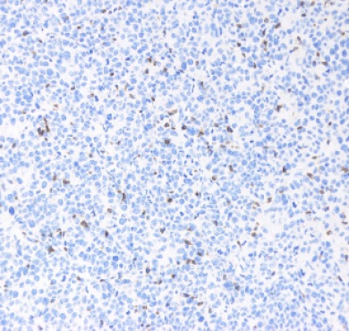
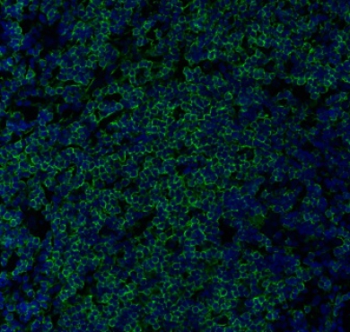
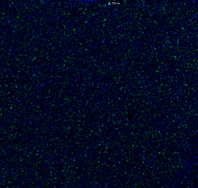
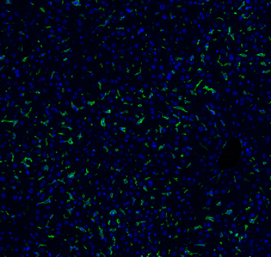
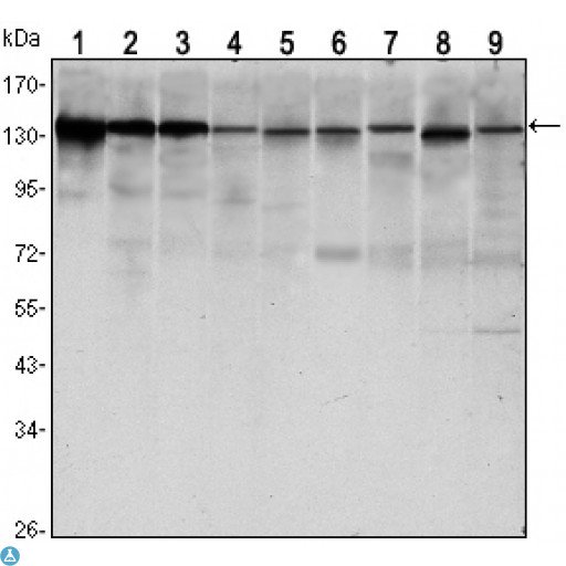
| Applications | ELISA, FC, IHC, WB |
| Dilution range | WB 1:500-1:2000 IHC 1:200-1:1000 FC 1:200-1:400 ELISA 1:10000 |
| Specificity | E-cadherin Monoclonal Antibody detects endogenous levels of E-cadherin protein. |
| Protein Name | Cadherin-1 CAM 120/80 Epithelial cadherin E-cadherin Uvomorulin CD antigen CD324 E-Cad/CTF1 E-Cad/CTF2 E-Cad/CTF3 |
| Immunogen | Purified recombinant fragment of human E-cadherin expressed in E. Coli. |
| Storage Instruction | Store at -20°C, and avoid repeat freeze-thaw cycles. |
| Note | For Research Use Only (RUO). |
| Validated Application |
| Host | Mouse |
|---|---|
| Clonality | Monoclonal |
| Clone ID | 7H12 |
| Reactivity | Human, Mouse, Simian |
| Conjugation | Unconjugated |
| Purification | Affinity purification |
| Isotype | IgGMale |
| Formulation | Ascitic fluid containing 0.03% sodium azide. |
| Gene ID | 999 |
|---|---|
| Gene Symbol | CDH1 |
| Database Links | HGNC:1748 OMIM:119580 |
| Alternative Names | Cadherin-1 CAM 120/80 Epithelial cadherin E-cadherin Uvomorulin CD antigen CD324 E-Cad/CTF1 E-Cad/CTF2 E-Cad/CTF3 |
| Function | Cadherins are calcium-dependent cell adhesion proteins . They preferentially interact with themselves in a homophilic manner in connecting cells; cadherins may thus contribute to the sorting of heterogeneous cell types. CDH1 is involved in mechanisms regulating cell-cell adhesions, mobility and proliferation of epithelial cells . Has a potent invasive suppressor role. It is a ligand for integrin alpha-E/beta-7. E-Cad/CTF2 promotes non-amyloidogenic degradation of Abeta precursors. Has a strong inhibitory effect on APP C99 and C83 production. |
| Post-translational Modifications | During apoptosis or with calcium influx, cleaved by a membrane-bound metalloproteinase (ADAM10), PS1/gamma-secretase and caspase-3 to produce fragments of about 38 kDa (E-CAD/CTF1), 33 kDa (E-CAD/CTF2) and 29 kDa (E-CAD/CTF3), respectively. Processing by the metalloproteinase, induced by calcium influx, causes disruption of cell-cell adhesion and the subsequent release of beta-catenin into the cytoplasm. The residual membrane-tethered cleavage product is rapidly degraded via an intracellular proteolytic pathway. Cleavage by caspase-3 releases the cytoplasmic tail resulting in disintegration of the actin microfilament system. The gamma-secretase-mediated cleavage promotes disassembly of adherens junctions. N-glycosylation at Asn-637 is essential for expression, folding and trafficking. Ubiquitinated by a SCF complex containing SKP2, which requires prior phosphorylation by CK1/CSNK1A1. Ubiquitinated by CBLL1/HAKAI, requires prior phosphorylation at Tyr-754. |
| Cellular Localization | Cell junction Cell membrane. Single-pass type I membrane protein. Endosome. Golgi apparatus, trans-Golgi network. Colocalizes with DLGAP5 at sites of cell-cell contact in intestinal epithelial cells. Anchored to actin microfilaments through association with alpha-, beta- and gamma-catenin. Sequential proteolysis induced by apoptosis or calcium influx, results in translocation from sites of cell-cell contact to the cytoplasm. Colocalizes with RAB11A endosomes during its transport from the Golgi apparatus to the plasma membrane. |
| Tissue Specificity | Non-neural epithelial tissues. |
| Swiss-Prot Key | P12830_HUMAN |
](/uploads/ueditor/20200825/1-200R5161100T7.png "Anti-E-cadherin antibody[STJ98009](图4)")